2025 AIChE Annual Meeting
(378e) Thiol-Based Coatings Improve Translucency of Elastomeric Anatomical Phantoms Fabricated By Vat Photopolymerization
Authors
Alan Aguirre-Soto - Presenter, Monterrey Institute of Technology and Higher Education
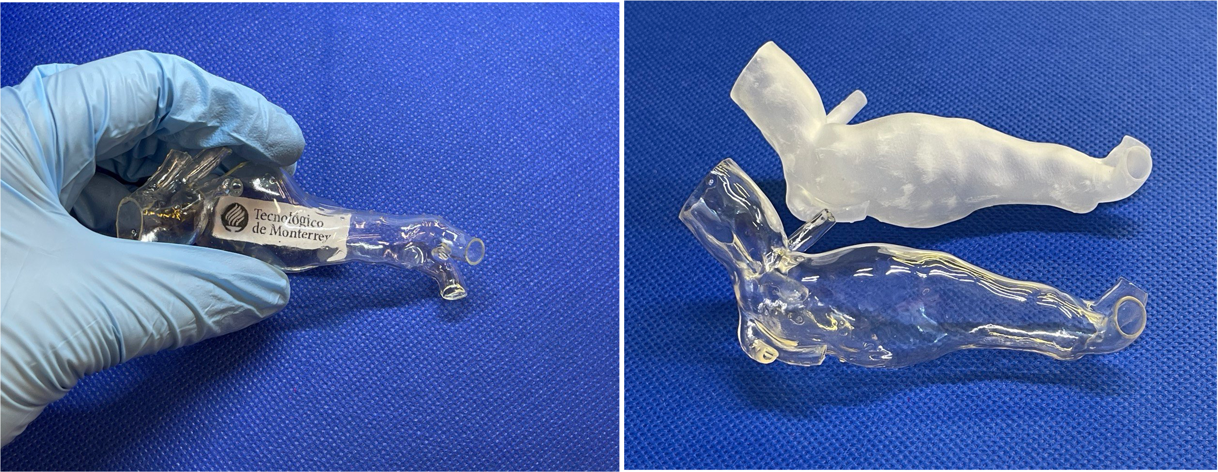
An ideal vascular phantom should be anatomically accurate, have mechanical properties as close as possible to the tissue, and be sufficiently transparent for ease of visualization. However, materials that enable the convergence of these characteristics have remained elusive. The primary goal of this research is to enable the creation of patient-specific 3D-printed phantoms with high anatomical accuracy. These phantoms hold immense potential for applications in pre-procedural planning and hemodynamic studies. We discuss the fabrication of patient-specific vascular phantoms with high anatomical fidelity, optical transparency, and mechanical properties close to those of vascular tissue. These final properties are achieved through the fabrication of patient-specific vascular models with vat photopolymerization on commercial elastomeric acrylic-based resins before coating them with thiol-ene formulations. A PETMP/allyl glycerol ether (AGE)/polyethylene glycol diacrylate (PEGDA) coating with a 30/70% AGE/PEGDA ratio applied on a flexible resin yielded elastic modulus, UTS, and elongation of 3.41 MPa, 1.76 MPa, and 63.2%, respectively, in range with the human aortic wall. The PETMP/AGE/PEGDA coating doubles the optical transmission from 40% to 80%, approaching the 88% obtained with the benchmark polydimethylsiloxane (PDMS). Higher transparency correlates with a decrease in surface roughness from 2000 to 90 nm after coating. Coated 3D-printed anatomical replicas are showcased for pre-procedural planning and medical training with good radio opacity and echogenicity. Thiol-click chemistry-based coatings, as a surface treatment for objects printed with vat photopolymerization techniques, address inherent limitations of photopolymer-based additive manufacturing (PAM). The presented approach can serve as a general post-processing technique for soft SLA 3D-printed parts to reduce the surface roughness to the nanoscale without compromising the material's flexibility. These accomplishments enable the previously unattainable goal of optical flow visualizations in soft patient-specific 3D-printed phantoms for laser Particle Image Velocimetry (PIV) studies, eliminating the reliance on conventional polymer casting methods. Furthermore, our approach allows for the precise replication of anatomical features with soft materials that closely mimic the mechanical properties of the vascular tissue, thus offering a promising alternative for next-generation vascular research and medical applications. These phantoms can be utilized both for Echo as well as for laser PIV. The techniques and materials developed herein have the potential to improve in vitro experiments in vascular research, precise simulations, and optical hemodynamic studies.