2025 AIChE Annual Meeting
Quantifying and Correcting Non-Uniform Deformation in Hydrogel Networks for Precision Expansion Microscopy
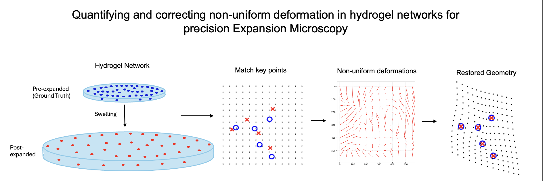
Expansion Microscopy (ExM) is a super-resolution imaging technique in which biological specimens are (1) embedded within a hydrogel and (2) swelled with water to cause its physical expansion. This process is typically assumed to produce isotropic expansion, preserving relative spatial relationships within the sample. In practice, however, heterogeneity in the gel’s polymer network and the biological specimen introduces local nonuniformities that can distort embedded nanostructures. To address this limitation, we developed a fiducial-based deformation mapping method to directly quantify and correct for non-uniform gel deformations. Protein-coated fluorescent microbeads are homogeneously dispersed throughout the hydrogel and imaged before and after expansion using fluorescence microscopy. The pre- and post-expansion images are then computationally registered, yielding both a precise global expansion factor and local displacement vector fields that capture deviations from ideal expansion. These vector fields are subsequently used to computationally restore the specimen’s original geometry, correcting distortions in the expanded micrograph. Altogether, this framework provides a quantitative and materials-informed correction strategy that enhances the accuracy and reliability of ExM-based nanoscale imaging.