2025 AIChE Annual Meeting
(603d) Interfacial Analysis of Brain Related Multi-Lipid Membranes Via Fluorescent Probe and Phase Diagram
Authors
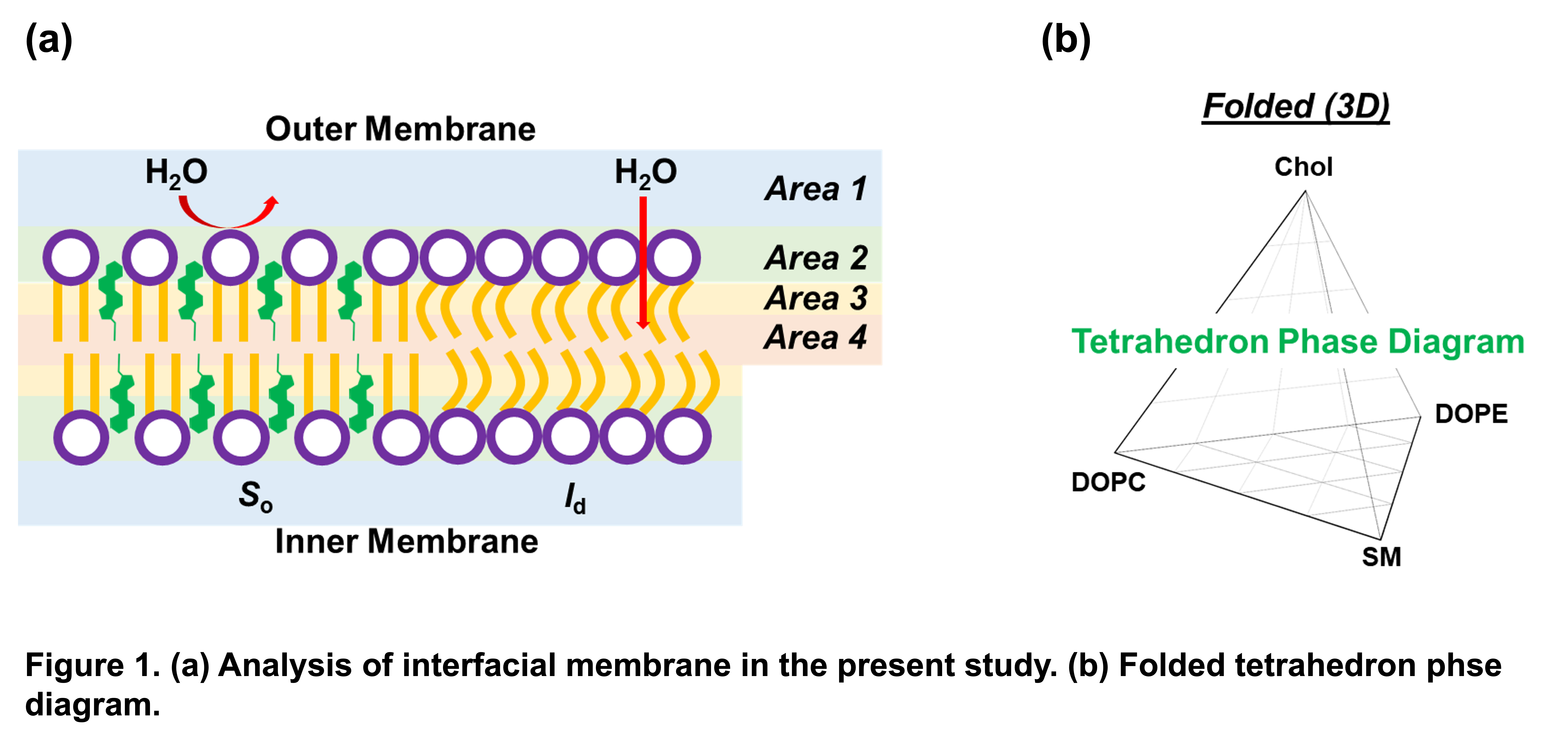
One method of analyzing the lipid membrane in the absence of other aspects of membrane relevant structures is via the use of small unilamellar vesicles (SUVs), also known as liposomes. Liposomes are commonly used as a biomimetic environment to study the biological membrane due to the simplicity of their structure alongside the ease of altering properties such as interfacial polarity and fluidity. Different lipid compositions of liposomes are able to be constructed that allows the researcher to study variations in characteristics such as polarity, fluidity, phase state, and hydrophobicity among others. By constructing liposomes with different compositions of brain membrane mimicking lipids, analysis may be performed on relevant brain membrane properties in isolation of proteins and other macromolecules present in a living neuron.
This study utilized porcine brain sphingomyelin (SM), 1,2-dioleoyl-sn-glycero-3-phosphocholine (DOPC), 1,2-dioleoyl-sn-glycero-3-phosphoethanolamine (DOPE), and cholesterol (Chol) as designated lipids to mimic commonly occurring brain lipid membranes. One of the largely utilized methods to analyze the properties of a membrane is the ternary phase diagram. Ternary diagrams are useful in that they are able to integrate 3 separate lipids at various ratios to display how altering the ratios may impact the polarity, fluidity, and ultimately the phase of the analyzed membrane. Although these diagrams are important additions to the field of lipid research, one of the most observed limitations is that only 3 lipid component membranes may be analyzed. Conventional phase diagrams are limited to displaying a maximum of only three components per diagram whereas the trends of more complex membrane systems are an important area of research that should also be thoroughly understood, as biologically relevant membranes have much more complex compositions. This work has the aim to introduce a 3D tetrahedron phase diagram that incorporates 4 separate ternary phase diagrams joined together at their edges to allow for the inclusion of an additional fourth lipid for use in research projects in lipid research.