Egfr Binding Peptide Contrast Agents for Signaling Egfr-Positive Tumors
2024 AIChE Annual Meeting
Egfr Binding Peptide Contrast Agents for Signaling Egfr-Positive Tumors
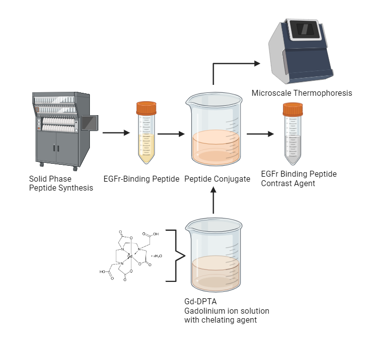
Triple Negative Breast Cancer, TNBC, is an aggressive and fast-growing subtype of Breast Cancer, which lacks an established therapeutic target. It is characterized by the absence of increased expression of the estrogen receptor, progesterone receptor, and human epidermal growth factor receptor 2 (HER2). Additionally, TNBC affects 13 in every 100,000 women in the United States. Magnetic Resonance Imaging, MRI, is the main noninvasive method to detect these tumors. However, due to TNBC’s unique characteristics and nonuniform shape, it is more likely to have inaccuracies in the diagnosis of the tumor and/or tumor morphology. A gadolinium-based contrast agent chelated by an Epidermal Growth Factor Receptor (EGFr) binding peptide is being developed to address these limitations in MRIs. The use of this conjugate will improve the contrast in MRI scans, leading to more accurate detections of EGFr-positive tumors, such as the most common subtypes of TNBC. The conjugate will bind to EGFr, where the paramagnetic gadolinium ions will interact with water molecules in tissues, influencing relaxation rates on MRI scans. EGFr-binding peptides were synthesized by Solid-Phase Peptide Synthesis and coupled with a chelating agent. The main peptide, AEGFr, was found in previous literature and was linked to self-assembling peptides previously developed in KumarLab to develop novel self-assembling multidomain peptides SAP1-AEGFr and SAP2-AEGFr. The peptide conjugates were synthesized and characterized for their chemical, structural, and mechanical properties. The affinity of self-assembling conjugated peptides to the EGFr needs to be further tested with Microscale Thermophoresis, MST to find the best binding affinity. Next, the peptide conjugates are combined with a gadolinium(III) ion solution. The peptide will then self-aggregate to form a translucent soft hydrogel. The formed EGFr Binding Peptide Contrast Agent can then be injected into a patient, increasing the accuracy of EGFr+ tumor diagnosis.
Figure 1: Schematic of Experimental Procedure. Created with BioRender.com