2024 AIChE Annual Meeting
(183e) Nanoscale Mechanical and Morphological Characterization of Ebolavirus-like Particles: Implications for Therapeutic Development
Authors
Ebolavirus (EBOV) is one type of filovirus that causes the deadly EBOV disease, with an average fatality rate of around 50%. EBOV outbreaks are devastating and unpredictable and may emerge as the next global pandemic. As a BSL-4 pathogen, EBOV is inaccessible to regular biological laboratories. Therefore, EBOV virus-like particles (EBOV-VLPs) and EBOV pseudoviruses (EBOV-PVs) are utilized in initial development of many potential therapies, as opposed to using live viruses. These particles must accurately represent the morphological, mechanical, and biochemical properties of live EBOVs. Due to EBOVs’ nanometer size and irregular, filamentous shape, these properties have been challenging to characterize. In this research, state-of-the-art nanoscale characterization techniques were employed to examine surface characteristics of a selection of commonly used EBOV-approximating particles. In summary, the study comprehensively determined the accuracy of EBOV approximation with virus-like particles and pseudovirus, and the uniformity of mechanical, structural, and biochemical traits across different EBOV approximations. This provides important implications for developing therapeutic treatments against EBOV using these approximating reagents.
- Methods
Atomic force microscopy (AFM) was used to characterize each of the EBOV-approximating particles. Each particle was observed hydrated under 1x phosphate buffer serum (PBS), to approximate the particles’ in-vivo state. During analysis, three protein solutions were used. The UV-inactivated EBOV (UV-EBOV) and the EBOV pseudovirus (PV) stock solutions were supplied by Wendy Maury at Iowa State University, and the EBOV-like particle (VLP) stock solution was supplied by IBT Bioservices. Each of these particles mimic membrane characteristics of EBOV, so they are well-suited to comparative study.
1.1. Peakforce AFM
Each particle was examined via Peakforce AFM imaging mode. This mode is owned by Bruker AFM and utilizes a variation of tapping mode which averages a certain number of force scans per pixel to construct a topographical image of the surface of the sample. By this method, individual nanoscale particles can be imaged topographically—this is how physical conformation of each particle was determined.
Another function of Peakforce AFM scanning is nanoindentation. By this method, force scans can be conducted precisely over the location of a particle based on location coordinates determined by a Peakforce AFM image scan. This force scan registers a small indentation in the surface of the particle, and by analyzing force and depth of the nanoindentation, elastic modulus of the particle can be established. To minimize interference from the substrate, nanoindentation depths of 1 to 5 nm were used in analysis. Any indentations deeper than this are assumed to be affected by the stiffness of the substrate and have been discarded from the dataset.
1.2. Adhesion Force Mapping
Adhesion force mapping is another force scanning AFM functionality, whereby an AFM cantilever is treated with an antibody particle, which is then contacted with a particle of interest adhered to a stiff surface. The force which is required to separate the tip from the surface is then analyzed to determine the binding affinity between the antibody and particle of interest. In this study, adhesion force measurements are taken over a single-particle sample, to distinguish between adhesion on the particle and adhesion over the surface. This allows negative control of each force map to be the substrate background, and sample adhesion can be thus compared to the negative control. As UV-EBOV is the closest approximation to live EBOV, adhesion force between UV-EBOV and antibody-treated tip will be regarded as the positive control for comparison between adhesion mapping of different particles. For specific sample preparation and testing protocols, see Supplemental Methods.
- Results
2.1. Ebola Virus-Like Particles
Although UV-EBOV and EBOV PV are known to be similar to live EBOV, neither is commercially available. For this reason, the EBOV-VLP is highly useful. The VLP possesses the outer membrane characteristics and some of the inner particle characteristics, but lacks the ability to self-replicate, making it safe for handling in a BSL-2 lab [1]. To determine whether this is a good approximation of live EBOV, the VLP was characterized by the same methods which the UV-EBOV and PV were characterized, and then compared to the UV-EBOV characterization as the most EBOV-like particle.
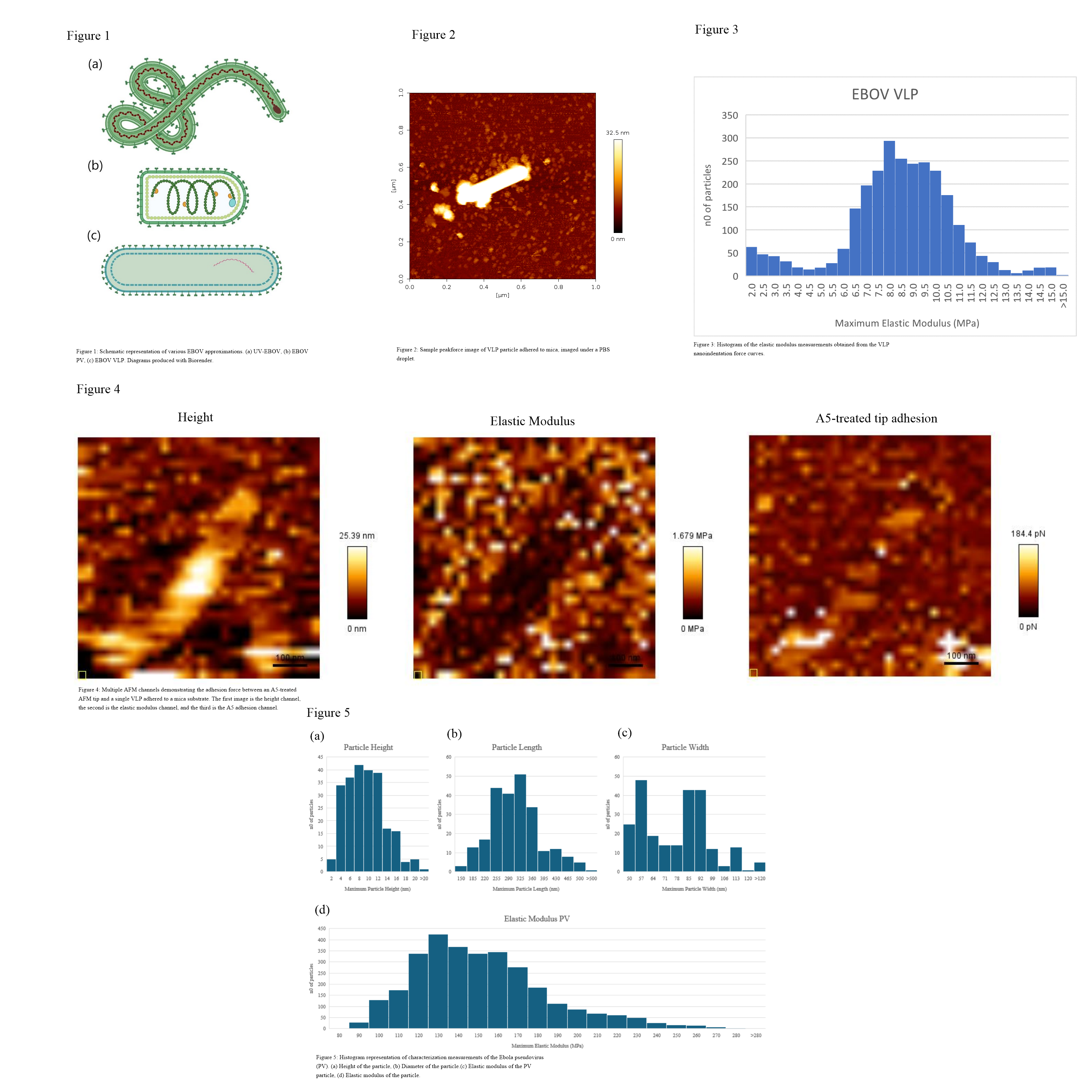
Peakforce AFM imaging of VLP particles on a treated mica surface revealed that the average width of the VLP particle when hydrated in PBS was 115.1 ± 55.5 nm, the average length of the particle was 480.2 ± 152 nm, and the average height was 51.5 ± 18.9 nm. Overall, the particles exhibited a cylindrical or pill shape. A sample Peakforce scan of the VLP particles adhered to a treated mica surface can be observed in Figure 2.
When hydrated in PBS, the VLP had an average elastic modulus of 8.54 ± 1.69 MPa. The histogram of elastic modulus data is depicted in Figure 3.
Another important consideration when characterizing the VLP particle is determining whether the particle has PS on the surface of the membrane, like live EBOV does. This was assessed using Adhesion Force Mapping, which characterized adhesion forces between a Bruker MLCT probe treated with Annexin V (A5), and the VLP. The resulting force map can be seen in Figure 4.
2.2. Ebola Pseudovirus
While the closest allegory to live EBOV is UV-EBOV, it is not always feasible for a group to obtain. At the time of writing, UV-EBOV is commercially unavailable, and must be obtained from a BSL-4 lab. If a lab cannot do this, another commonly used approximation of EBOV is Ebola pseudovirus (PV), which is formed by expressing EBOV glycoproteins on the surface of a more benign virus. To assess EBOV PV as an approximating particle, our lab obtained a sample from our collaborator of vesicular stomatitis virus (VSV) which has had EBOV GP artificially implanted in the surface of the virus to mimic EBOV’s ability to enter the endosome. The PV was then tested and in the coming weeks will be analyzed against the characteristics of UV-EBOV.
When characterizing the PV, AFM Peakforce tapping mode revealed that the PV particles exhibited an oblong pill shape, with a domed top. The average width of the particle was 70.4±20.3 nm, the average length was 285±74.6 nm and the average maximum height of the particle was 7.4±4.2 nm. Figure 5 parts (a), (b) and (c) demonstrate the distribution of height, length, and width measurements of individual particles, respectively. Peakforce nanoindentation of a PV-saturated mica substrate revealed that the average elastic modulus of the PV particle was 0.64 MPa, with a median of 0.49 MPa, a geometric mean of 0.47 MPa, and a standard deviation of 0.51 MPa. Figure 5 part (d) depicts the distribution of elastic modulus measurements across several hundred force curves. It is of note that while the histogram has a left-leaning distribution, there is a distinct peak and then fall to the histogram data, which indicates that the peak of the histogram is interpretable, and the elasticity is large enough to be measured by Peakforce nanoindentation methods.
To assess the presence of phosphatidylserine (PS) on the EBOV PV surface, adhesion force mapping can be conducted between an AFM cantilever tip treated with Annexin V (A5) and a mica substrate saturated with PV particles. As A5 is a known antibody of PS, this method demonstrates the presence of PS on the particle surface by showing the tip’s adhesion to the particle. At the time of abstract submission, this work has not yet been completed, but this will be carried out imminently. Additionally, the method has been proven to work with EBOV-VLP samples.
2.3. UV-Inactivated Ebolavirus
Of the three particles of interest being examined, the sample which is assumed to be most like live Ebolavirus is the UV-inactivated Ebolavirus particles. The only difference between these particles and live EBOV is that the replicative proteins at the nucleus of the virus have been irradiated with UV light, such that the particle is not capable of self-propagating. AFM testing has yet to be conducted on this approximating particle, but from literature information it is expected that UV-EBOV has a long, filamentous structure roughly 50 nm wide and high, and approximately 1000 nm long, and an elastic modulus of the particles of approximately 4-6 MPa. Work to confirm this will be conducted over the next several weeks.
- Conclusions
The characterization of ebolavirus-approximating particles is critical to the ongoing development of therapeutic drugs to combat the deadly EBOV disease. Overall, VLP demonstrated a physical configuration and elasticity more aligned with the filamentous shape of EBOV and the known stiffness of other Marburg viruses, while PV had a rounder, softer configuration. Additionally, each approximating particle has EBOV GP and PS expressed on the surface. This combined with the fact that VLP is available to the consumer, while PV is not, makes VLP the more attractive approximation for EBOV when conducting experiments regarding therapeutic drug development.
